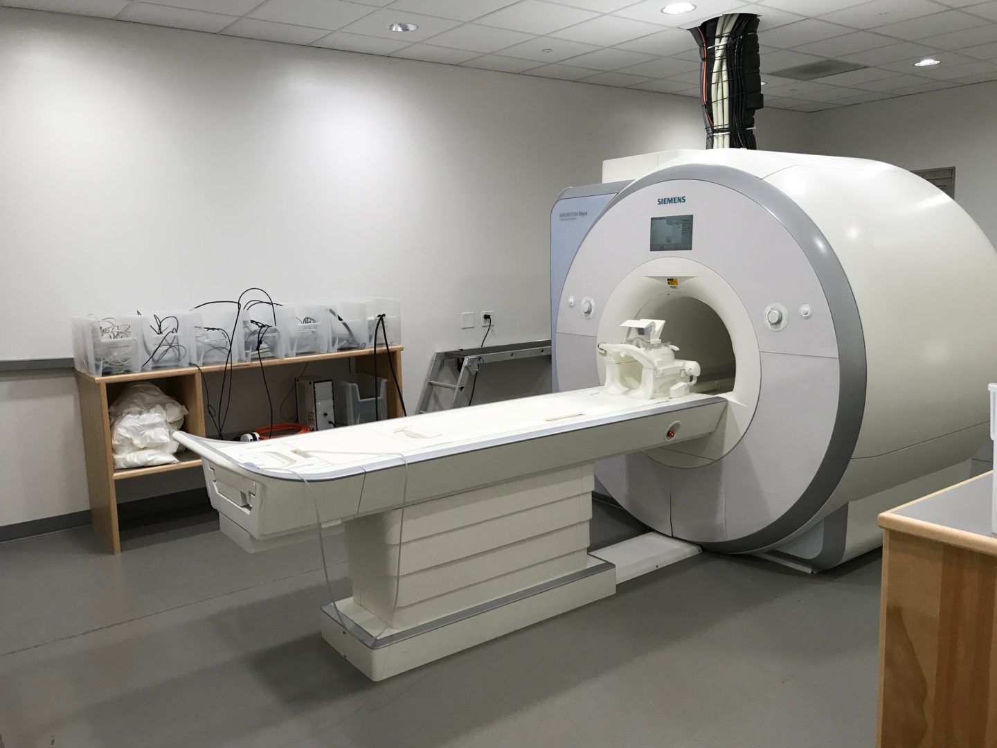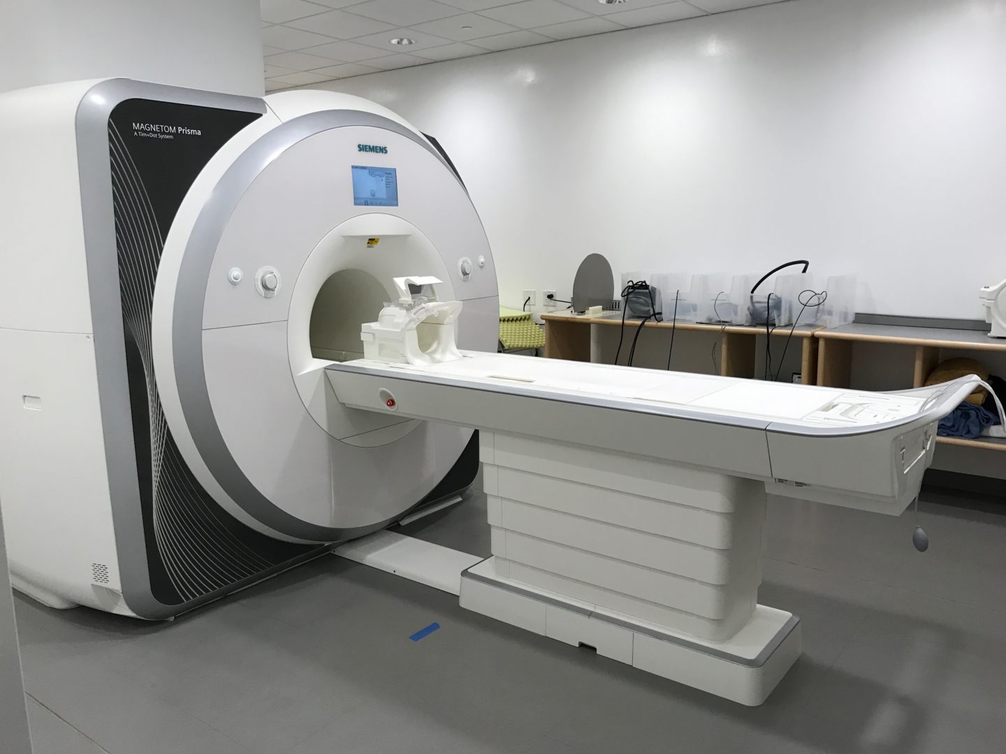
For over three decades, functional MRI has allowed researchers to use changes in blood oxygenation as a proxy for mental activity, allowing them to peer into the thinking brain safely and non-invasively with unprecedented spatial precision. Our MRI Facility is equipped with two research-dedicated 3 Tesla MRI scanners (a Siemens 3T MAGNETOM Skyra and a 3T MAGNETOM Prisma). The scanners are identically equipped with all the hardware and software necessary to perform brain imaging. The scanners can be run independently or synced to scan two participants in a coordinated task in what has become known as “hyperscanning”. For those studying special populations such as children, the MRI facility offers a simulator that provides a realistic approximation of an actual MRI scanner to allow acclimation and training in a controlled setting.
Technical details
The Skyra scanner was installed in late 2010 and became fully operational in early 2011. The Prisma scanner was installed in mid-2014 and became fully operational in late 2014. Images may be acquired with either a 20- or 64-channel phased array coil. The Prisma scanner features a 60 cm bore with very strong 80 mT/m @ 200 T/m/s gradients and supports up to 128 channels, while the Skyra scanner features a larger 70 cm bore with 45 mT/m @ 200 T/m/s gradients and supports up to 64 channels. Product Siemens pulse sequences for structural imaging, as well as multi-band-capable BOLD and diffusion EPI sequences are provided.
Both magnet rooms are equipped with MRI compatible behavioral equipment. A PST projector displays visual stimuli. Subject responses may be collected using 2 different types of button box systems (PST and Current Designs). Auditory stimuli may be delivered to the subject using an Avotec Silent Scan system. Subjects’ verbal responses can also be collected via a noise canceling microphone (Optoacoustics FOMRI III Mic). Each scanner has a Eyelink 1000 (SR Research) mounted in bore for high speed eye tracking.
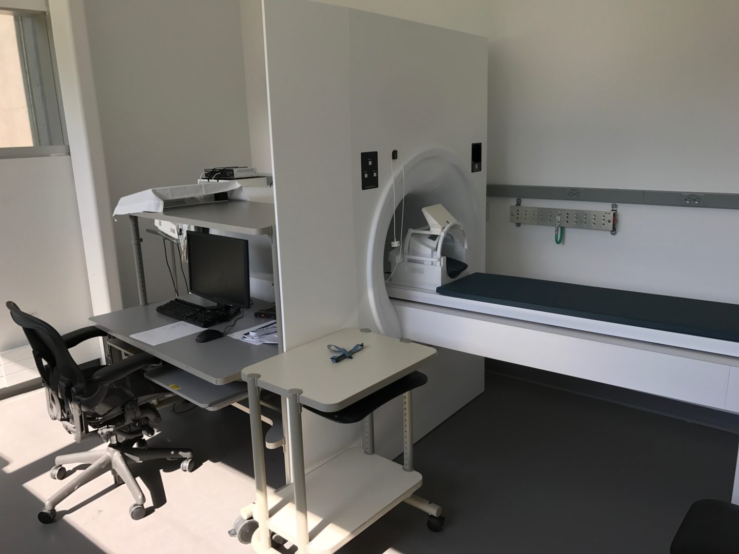
MRI Simulator
The Scully Center houses a PST MRI simulator for researchers who would benefit from acclimation sessions with their participants (patient populations, pediatric populations). The simulator provides a realistic approximation of an actual MRI scanner to allow acclimation and training in a controlled environment for a fraction of the cost of MRI access. The simulator is equipped with a realistic mock head coil, mechanical bed, speakers, and a library of the MRI scanner sounds to help acclimate the subject to an MR environment.
The simulator is also equipped with a MoTrack motion tracking system to help subjects train to keep their head still during experiments. Visual stimuli can be presented to the subject via a 1080p monitor at the back of the bore, and behavioral data can be recorded using X-Keys response boxes. The simulator is also equipped with an in-bore microphone to record subjects’ verbal responses and headphones for auditory stimuli.
PNI researchers interested in obtaining more detailed information regarding our MRI systems and peripheral equipment should visit our internal Scully Facilities Wiki.